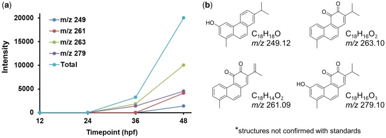
Fig. 4.
a) Signal intensity of metabolites identified by LC-MS in extracts from retene exposed embryos at 12, 24, 36, and 48 hpf. Each point represents average ion intensity from at least 3 replicate extracts from 36 fish each. b) Potential structures for metabolites corresponding to observed m/z ions.

