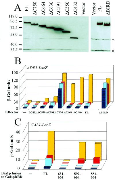
Figure 2.
Bas1p contains a small domain (BIRD) with interaction and regulatory functions. (A) Expression level of the deletion mutants of Bas1p in the bas1bas2 yeast strain. The bas1bas2 (L4233) strain was transformed with the empty control plasmid (vector) or YPL-BAS1 plasmids carrying the full-length (FL) or the deletion version of the BAS1 gene. Total yeast protein extracts were prepared as described in Materials and Methods. Proteins were subjected to SDS–PAGE and electroblotted on a polyvinylidene difluoride membrane. The blot was then incubated with anti-Bas1p purified antibodies (1 µg IgG/ml) followed by horseradish peroxidase-labeled IgG (1/6000 dilution) as secondary antibodies and, finally, with luminescent substrate before exposure to film. The cross-reacting bands are marked by asterisks. Comparable levels of the different Bas1p proteins were also observed in a gcn4bas1BAS2 strain (not shown). (B) In vivo effects of the deletions in Bas1p assayed with the ADE1–LacZ reporter. The bas1BAS2 (Y329, yellow and red boxes) and bas1bas2 (L4233, light and dark blue boxes) cells were co-transformed with a plasmid carrying the ADE1–LacZ fusion and the different YPL-BAS1 plasmids carrying the full-length (FL) or deleted BAS1 gene. The lane ‘Effector –’ corresponds to transformation of yeast strains with the centromeric vector not carrying the BAS1 gene (YPL). Transformed cells were grown in the presence (red and dark blue boxes) or absence (yellow or light blue boxes) of adenine and β-Gal assays were performed as described in Materials and Methods. (C) In vivo role of BIRD in the Bas1p–Bas2p interaction monitored by a two-hybrid approach. The Y187 yeast strain containing an ADE2-expressing plasmid (pAZ11) (27) was transformed with a centromeric bait plasmid expressing Gal4p DBD fusions (pDBT) (16) and with a second prey plasmid expressing (yellow and red boxes) or not (light and dark blue boxes) the Bas2p–VP16 chimera. Cells were grown in the presence (red and dark blue boxes) or absence (yellow and light blue boxes) of adenine in SC medium lacking histidine and tryptophan. β-Gal assays were performed as described in Materials and Methods.

