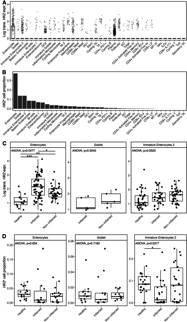
Fig. 5.
HK2 is mainly expressed by mature enterocytes and increased during inflammation. HK2 expression was analyzed in published single-cell RNA sequencing data derived from mucosal biopsies of UC patients [18]. A HK2 expression per cell type. Box plot depicting the median and 25th–75th percentile. Whiskers indicate most extreme points within 1.5-times interquartile range deviance from the median and dots represent samples outside of this interval. Note that only enterocytes express significant HK2 levels; thus, only here an interval box is visible. B Abundances of cell types expressing HK2. C HK2 expression in mature and immature enterocytes and goblet cells divided into samples from healthy controls, inflamed and non-inflamed mucosa. D Abundances of cell types expressing HK2 after stratification into disease group. ANOVA p values denote whether significant differences among the three groups exist that were then further analyzed by pairwise Wilcoxon tests (*p < 0.05, **p < 0.01, ***p < 0.001)

