Figure 3.
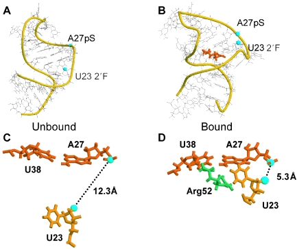
(A and B) Conformational changes in TAR upon tat peptide binding. The phosphate backbone is represented by yellow ribbons, highlighting rearrangements of the bulge residues generated by peptide binding. Blue spheres denote 19F U23 and pS A27 label positions used in the present work. Illustrations and distances shown are adapted from solution NMR-derived models of the free and peptide-bound RNA (5,21). (A) Unbound conformation (PDB #1ANR, model one). (B) Bound conformation (PDB #1ARJ, model one.) Tat residue arg52, which provides key contacts with the RNA, is shown in orange. The RNA has straightened from the unbound conformation, residues C24 and U25 are looped out of the helix, and residue U23 has shifted to a new position adjacent to G26 and A27. (C and D) Rearrangements at TAR bulge binding site upon tat peptide binding. Blue spheres again denote 19F U23 and pS A27 label sites. (C) Close-up of unbound conformation, showing relative positions of U23, A27 and U38, and the inter-label separation monitored in the present study (PDB #1ANR, model one; 19F-pS inter-label separation is 12.3 Å in this model). (D) Close-up of bound conformation. Tat residue arg52 is shown in green. TAR accommodation of the tat peptide results in a significant repositioning of U23, drawing it into close proximity to A27 and U38, accompanied by a large decrease in separation between the 19F U23 and pS A27 label positions (PDB #1ARJ, model one.) Following binding, the 19F–pS distance has decreased to 5.3 Å in the model shown.

