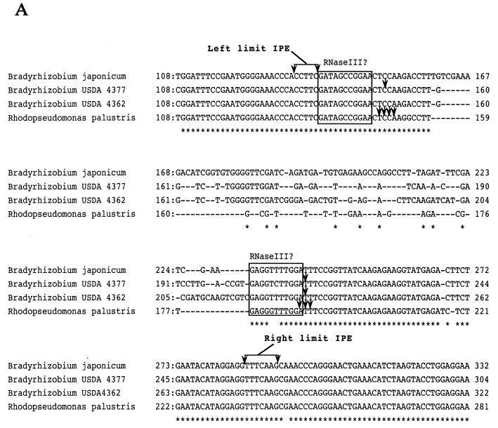
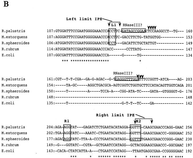
Figure 2.
(A) Sequence alignment of internal DNA segments from Bradyrhizobial 23S rRNAs cloned by PCR. The origins of the sequences are as indicated on the left side of each line of sequence and the positions within 23S rRNA are shown at the beginning and end of each line. The numbering of bases for Bradyrhizobium USDA 4362 and USDA 4377 23S rRNAs is arbitrary since complete sequences have not been determined in these cases. Identical bases are marked (*). Arrowheads indicate positions of RNase III cleavage sites determined here and from studies with R.palustris (20). In each case, adjacent partially homologous sequences of 11 nt on the left of the RNase III cleavage sites are boxed to indicate potential binding sites for RNase III. On the top and bottom, sets of sequence arrows bracket the sequence representing the range of endpoints (left and right limit IPE) found in the in vivo RNA as determined in this and the previous study. (B) Sequence alignment of internal segments of 23S rRNAs from E.coli and representative α-proteobacteria containing and lacking 5′ IVSs. The numbering for M.extorquens 23S rRNA is arbitrary since a complete sequence has not been determined in this case. Limits of the IPE as determined from studies with R.palustris are shown by brackets and the putative RNase III binding sites are boxed. Also boxed are sequences denoted as L1, R1 and R2 where homology occurs in M.extorquens, R.palustris and R.sphaeroides but not the other species. Arrowheads indicate RNase III cleavage sites determined by primer extension.


