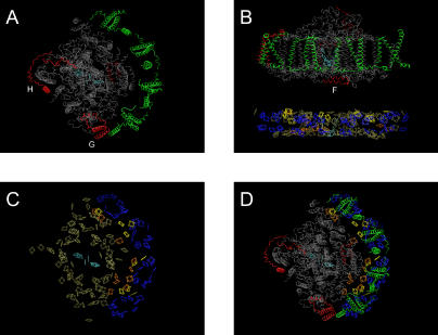
Figure 5. Crystal Structure of PS-I as Determined by Ben-Shem et al. [7,53], Visualized in VMD [52], and Rendered with POV-Ray 3.6.
The color scheme is adapted from Ben-Shem et al. [7,53] and is interpreted as follows. Portions shown in gray are common subunits found in both PS-I reaction centers in cyanobacteria and higher plants, while the red subunits are unique to higher plants. The two LHCI heterodimers are shown in green. The two different orientations of selected PS-I complex are shown top-down (A) and in a side view (B). In (C) and (D) the chlorophyll molecules are colored by a putative function: the P700 core antennae are tan, the special pair (P700) and voyeur chlorophyll molecules are cyan, the linker chlorophyll between the LHCI heterodimers is in yellow, the antennae chlorophyll molecules complexed within an LHCI subunit are in blue, and the “gap” chlorophyll molecules are shown in orange. (A), (C), and (D) are views looking down on the stromal hump highlighting protein, chlorophyll, and composite, respectively. (B) faces the LHC belt from inside the plane of the thylakoid membrane; only protein is shown in the upper portion and only chlorophyll below.

