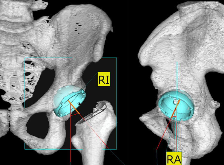
Figure 1. Radiographic visualization of acetabular component orientation.
The left image depicts the acetabular component orientation, with the RI measured to evaluate the angle of the cup's tilt in the coronal plane. The right image displays the RA, assessing the cup's rotation angle in the axial plane.
RA, radiographic anteversion; RI, radiographic inclination

