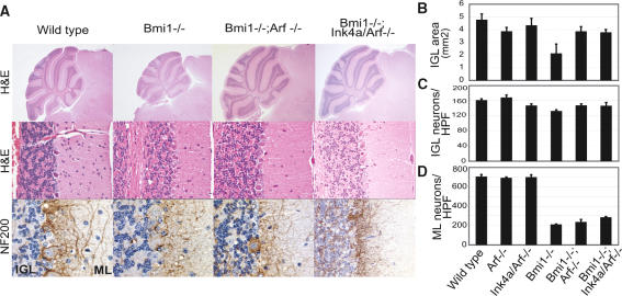
Figure 3.
Ink4a and Arf differentially contribute to histological abnormalities of the Bmi1-/- cerebellum. (A) Morphological analysis of wild-type (left panels), Bmi1-/- (middle left panels), Bmi1-/-;Arf-/- (middle right panels), and Bmi1-/-;Ink4a/Arf-/- (right panels) adult cerebellum. Haematoxylin and eosin (H&E, top and middle panels) staining shows rescue of overall cerebellar size and granular layer thickness in Bmi1-/-;(Ink4a)Arf-/- mice. (Bottom panels) Aberrant arborization of basket neurons (NF200 staining) is observed in all cerebella lacking Bmi1. Final magnification, 5× and 60×. (IGL) Internal granular layer; (ML) molecular layer. (B) IGL area measurements reveal similar significant rescues in Bmi1-/-;Arf-/- and Bmi1-/-;Ink4a/Arf-/- mice. (C) Ink4a/Arf loss induces a complete rescue of the number of Bmi1-/- granule neurons, whereas Arf loss leads to a partial rescue (p < 0.01). (D) The partial rescue in number of Bmi1-/- molecular layer neurons is significantly better in an Ink4a/Arf deficient background (p < 0.05) (HPF, high-power field; n = 4 mice).

