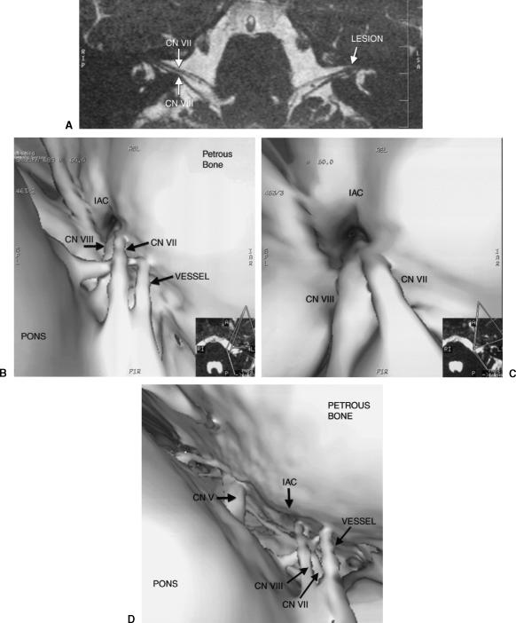
Figure 1.
(A) Oblique axial reformat of the CISS data showing CNs VII and VIII bilaterally. The tiny intracanalicular lesion near the fundus of the left IAC is likely an acoustic neuroma. (B) Surface model of CISS data showing CNs VII and VIII in the CPA with a vessel passing between them. (C) Closer view from the 3D navigational program showing the porus acusticus with CNs VII and VIII. (D) Surface model displaying the surgical view of the CPA cistern showing CNs V, VII, and VIII. CISS, constructive interference in the steady state; CNs, cranial nerves; IAC, internal auditory canal; CPA, cerebellopontine angle.

