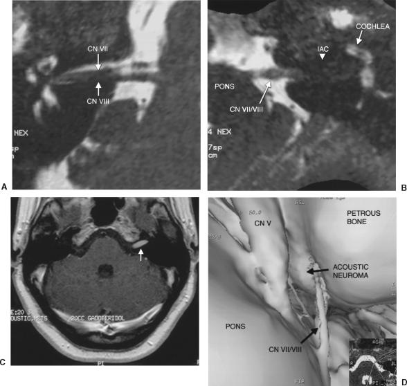
Figure 3.
(A) Oblique axial reformat of CISS data shows the right IAC with CNs VII and VIII. (B) Oblique axial reformat of CISS data shows the left CPA with the acoustic neuroma filling the IAC. (C) Axial T1-weighted postgadolinium image shows an enhancing lesion in the left IAC consistent with an acoustic neuroma. (D) Surface-rendered model of the left petrous face shows the acoustic neuroma filling the IAC as CNs VII and VIII enter the canal. CISS, constructive interference in the steady state; IAC, internal auditory canal; CNs, cranial nerves; CPA, cerebellopontine angle.

