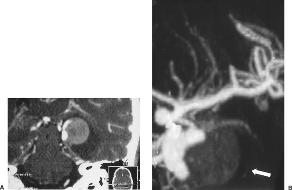
Figure 7.
(A) Reconstructed coronal CTA showing the aneurysm arising from the P2-P3 segment of the left posterior cerebral artery with a wide base and dissecting thrombus. (B) Reconstructed 3D maximum intensity pixel projection confirms the presence of the thrombus within the aneurysm (arrow). CTA, computed tomographic angiography.

