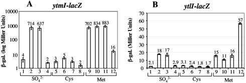
FIG. 3.
Expression of ytmI- and ytlI-lacZ fusions in wild-type, ytlI, spx, and rpoAcxs-1 strains. Cells of each fusion-bearing strain were grown in TSS media containing sulfate, cysteine, or methionine as the sole sulfur source. Samples were collected at late log phase, where maximum β-galactosidase activity was observed. The samples were assayed for β-galactosidase activity, which was expressed in Miller units (27). Experiments were performed in triplicate. (A) Bar graph of β-galactosidase activity in cells bearing the ytmI-lacZ fusion. Lanes 1, 5, and 9, strain ORB4794 (ytmI-lacZ); lanes. 2, 6, and 10, ORB4801 (ytmI-lacZ rpoAcxs-1); lanes 3, 7, and 11, ORB4802 (ytmI-lacZ spx); lanes 4, 8, and 12, ORB4804 (ytmI-lacZ ytlI). (B) Bar graph of β-galactosidase activity in cells bearing the ytlI-lacZ fusion. Lanes 1, 5, and 9, ORB4866 (ytlI-lacZ); lanes 2, 6, and 10, ORB4870 (ytlI-lacZ rpoAcxs-1); lanes 3, 7, and 11, ORB4871 (ytlI-lacZ spx); lanes 4, 8, and 12, ORB4873 (ytl-lacZ ytlI).

