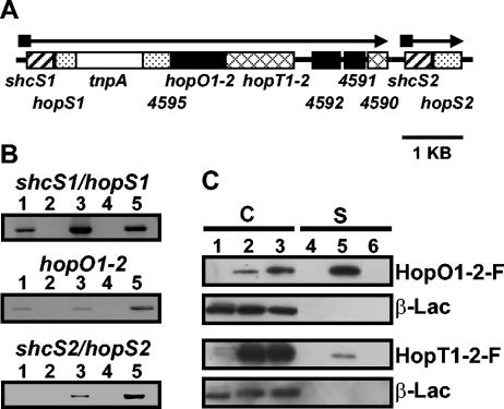
FIG. 2.
RT-PCR experiments indicate that shcS1/hopS1 and shcS2/hopS2 are transcribed in a type III-dependent manner and that HopO1-2 and HopT1-2 are both secreted in culture via the DC3000 TTSS. (A) The genetic organization of a cluster of genes in the DC3000 chromosome which are homologous to shcO1, hopO1-1, or hopT1-2. The homologous TTC genes, shcS1 and shcS2, are depicted with striped boxes. The homologous effector genes hopS1 and hopS2 are depicted with stippled boxes. Black boxes depict genes or ORFs that are homologous to hopO1-2 and hatched boxes depict hopT1-2 and a nearby homologous ORF, PSPTO4590. The open box represents a transposase that interrupts hopS1 and PSPTO4595. The ORFs with number designations represent the PSPTO numbers from the databases. The arrows show the predicted orientation of transcription and the black squares indicate the presence of hrp boxes, which are often found associated with type III-related promoters. (B) RT-PCR analysis of the DNA region shown in panel A. RNA was isolated from DC3000 cultures grown in either rich KB medium or hrp-inducing minimal medium. RT-PCR using RNA from KB-grown cultures with reverse transcriptase (RT) (lane 1) or without RT (lane 2); RT-PCR using RNA from hrp-inducing grown cultures with RT (lane 3) or without RT (lane 4); PCR using DC3000 genomic DNA (lane 5). The primer sets used were complementary to sequences within the coding regions of shcS1/hopS1, hopO1-2, or shcS2/hopS2. (C) Immunoblot analyses were performed cultures of DC3000 or a DC3000 hrcC mutant disabled in type III secretion, carrying either pLN521 (encoding HopO1-2-FLAG) or pLN567 (encoding HopT1-2-FLAG). Cultures were separated into cell-bound (C, lanes 1 to 3) or supernatant (S, lanes 4 to 6) fractions. Lanes 1 and 4, wild-type DC3000; lanes 2 and 5, DC3000(pLN521) (top two blots) or DC3000(pLN567) (bottom two blots); lanes 3 and 6, DC3000 hrcC(pLN521) (top two blots) or DC3000 hrcC(pLN567) (bottom two blots). Strains also carried pCPP2318, which encoded a mature form of β-lactamase that should remain cell bound unless significant nonspecific cell lysis occurred. Cell-bound and supernatant fractions were concentrated 13.3- and 133-fold, respectively. Anti-FLAG antibodies were used to visualize HopO1-2-F and HopT1-2-F and β-lactamase (β-Lac) was detected with anti-β-lactamase antibodies.

