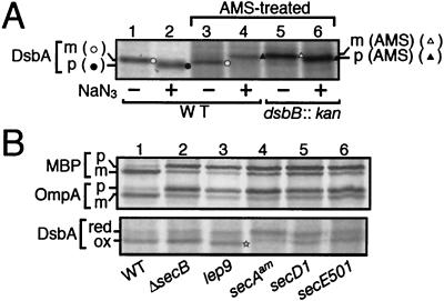
FIG. 2.
Redox states of newly synthesized DsbA as indications of its localization. (A) Migration of internalized and exported DsbA after AMS treatment. Cells of MC4100 (wild-type; lanes 1 to 4), and SS141 (dsbB::kan; lanes 5 and 6) were grown at 30°C and pulse-labeled with [35S]methionine for 60 seconds with (lanes 2, 4, and 6) or without (lanes 1, 3, and 5) prior 60-second exposure to 0.02% NaN3. Samples for lanes 3 to 6 were treated with AMS. Labeled proteins were precipitated with trichloroacetic acid, immunoprecipitated with anti-DsbA, and analyzed by SDS-PAGE. Open circles, unmodified mature form; solid circles, unmodified (oxidized) precursor form; solid triangles, AMS-modified (reduced) precursor form; open triangles, AMS-modified (reduced) and signal sequence-processed form. (B) DsbA synthesized in different mutant cells. Cells of TY0 (wild type; lane 1), CK1953 (ΔsecB; lane 2), IT41 (lep9; lane 3), MM66 [secA(Am); lane 4], THE521 (secD1; lane 5), and PR520 (secE501; lane 6) were grown at 30°C and pulse-labeled with [35S]methionine for 60 seconds. Samples for the upper panel were directly immunoprecipitated with anti-MBP and anti-OmpA, whereas those for the lower panel were first treated with AMS and then precipitated with anti-DsbA. The precursor and mature forms of MBP and OmpA are indicated by p and m, respectively. The reduced and oxidized forms of DsbA are indicated by red and ox, respectively. The oxidized but signal sequence-retaining DsbA species in the lep9 mutant is marked by a star.

