Figure 5.
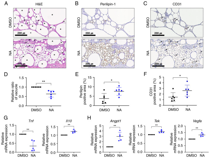
NA functions in fat engraftment by improving inflammation and angiogenesis. (A) H&E staining within grafts treated with DMSO or NA. The black asterisks indicate formed vacuoles or oil cysts within the grafts and the black arrows indicate the vessels within the grafts. Scale bars: 200 μm. Immunohistochemistry analysis of (B) perilipin-1 and (C) CD31 within grafts treated with DMSO or NA. Black arrows indicate the vessel structures within the grafts. Scale bars: 200 μm. The proportion of (D) vacuole area, (E) perilipin-1+ area and (F) CD31+ area measured within the whole grafts. (G) RT-qPCR analysis of Tnf and Il10. (H) RT-qPCR analysis of Angpt1, Tek and Vegfa. Data are presented as the mean ± SD. Statistical significance was assessed using unpaired Student's t-test: *P<0.05 and **P<0.01. DMSO, dimethyl sulfoxide; H&E, hematoxylin and eosin; NA, nervonic acid; RT-qPCR, reverse transcription-quantitative PCR.

