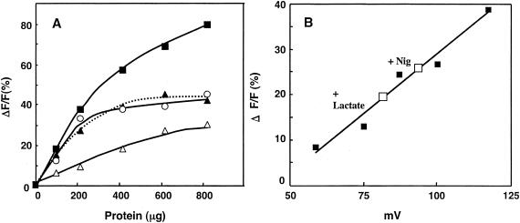
FIG. 2.
Membrane potential generation with different respiratory substrates and quantification of lactate-dependent membrane potential in ISO membrane vesicles. (A) The reaction mixtures contained different concentrations of ISO membrane vesicles in 50 mM KPB (pH 6.5) containing 5 mM MgSO4 and 2 μM diBA-C2-(5). The reactions were started by the addition of 10 mM lactate (▪), 10 mM succinate (▴), 0.5 mM NADH (○), or 3 mM ATP (pH 7.0) (▵), followed by the addition of nigericin (Nig) and valinomycin. ΔF/F indicates the rate (%) of fluorescence quenched per total unit of fluorescence. (B) ISO membrane vesicles were washed twice with 50 mM sodium phosphate buffer (NaPB, pH 6.5) to replace KPB with NaPB. The ISO membrane vesicles (10 μl; 101 μg of protein) were used for valinomycin-induced potassium diffusion potential measurement (▪), as described in Materials and Methods, and also for lactate-dependent Δψ (□) without Nig (+Lactate) or with Nig (+Nig) in 1 ml of the same buffer system as A.

