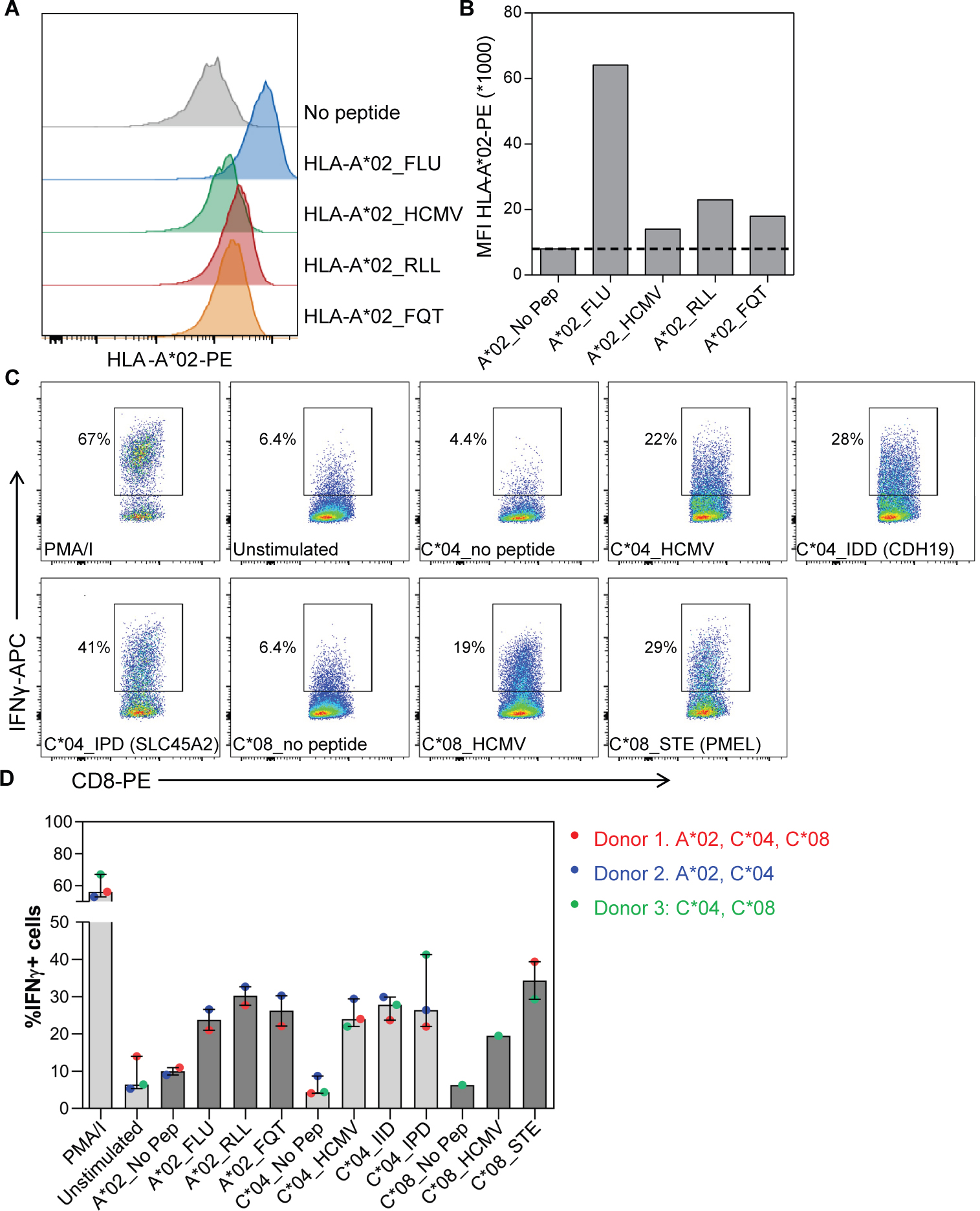
Figure 5. Shared splicing neoantigens bind HLA and induce T-cell reactivity.

(A) Histograms and (B) graph show HLA-A*02-PE staining on HLA-A*02 containing TAP deficient T2 cells without peptide (no pep), loaded with FLU and HCMV control peptides and RLLGTEFQT (RLL) and FQTTRRAMTL (FQT) peptide neoantigens. MFI = median fluorescence intensity. PE = Phycoerythrin conjugated antibodies. C) Dot plots and (D) graph show the percentage of Interferon gamma-positive (IFNγ+) CD8+ T-cells in response to 5 melanoma shared splicing antigens compared to negative (unstimulated, no pep) or positive (PMA/I, FLU, HMCV) controls. CD8+ T cells were primed using peptide loaded monocyte derived dendritic cells and thereafter tested against 721.221 cells selectively expressing the indicated HLA allele with and without peptide loading. Bars indicate median of 2–3 donors and lines interquartile range.
