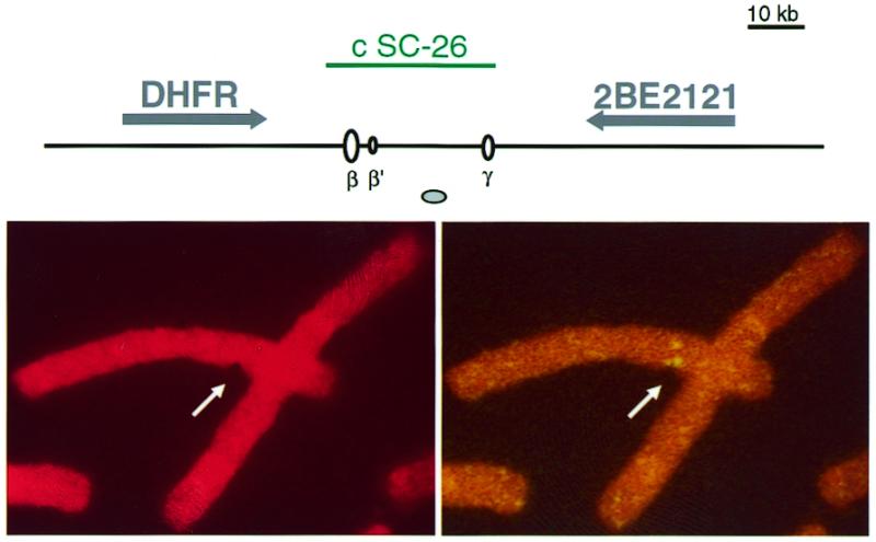
Figure 5.

Example of an aphidicolin-induced break at the Chinese hamster DHFR locus. (Top) Map of the analysed region. The cosmid probe used in this study, genes, DNA replication origins and MAR are represented as in Figure 1. (Bottom) A split signal colocalises with an aphidicolin-induced chromatid break at the DHFR locus. (Left) Propidium iodide staining. (Right) FISH with a biotinylated c SC-26 probe.
