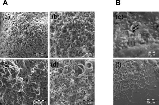
FIG. 2.
Morphological differentiation of S. coelicolor A3(2) grown on glass beads. (A) Image a shows a young colony composed of only substrate mycelia. Later on, aerial mycelia started to stand up into the air (b) and elongate (c), coincident with the appearance of pigments. Later, coiled aerial mycelia started to form septa and spores (d). Images a, b, c, and d were taken at 6, 16, 19, and 36 h after transfer to glass beads, respectively. The magnified image in d shows an aerial mycelium septated to form spores. Panel B shows spores germinating on glass beads after 8 h (e) and primary network of mycelium formed after 24 h (f). One or two germ tubes coming from the spore were observed (arrows).

