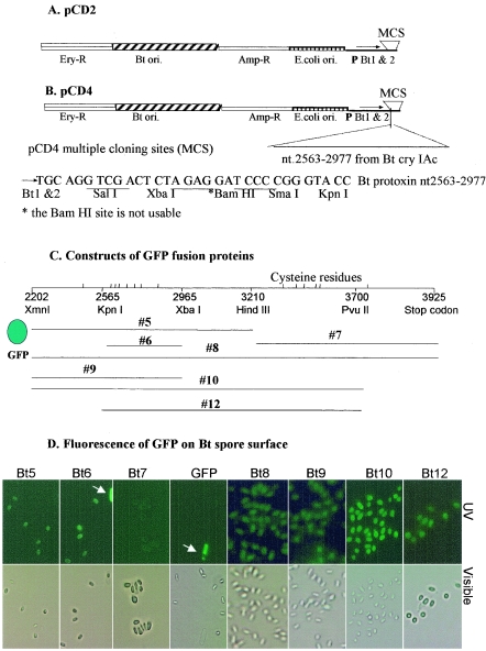
FIG. 1.
Display of GFP on the B. thuringiensis spore surface. (A) Vector pCD2 (6.6 kb) was derived from pHT3101 by inserting the B. thuringiensis toxin promoter region into the PstI site. This plasmid can grow in both E. coli (with ampicillin selection [Amp-R]) and B. thuringiensis (with erythromycin selection [Ery-R]). The inserted promoters (P) are sporulation specific. (B) pCD4, derived from pCD2, is a B. thuringiensis spore surface display vector. A 415-bp fragment from the cry1Ac gene was inserted downstream of multiple cloning sites. Fusion proteins can be displayed on the B. thuringiensis spore surface anchored by the portion derived from the B. thuringiensis protoxin. (C) GFP fusion proteins are shown schematically. The approximate nucleotide numbers and restriction sites are shown. The positions of cysteine residues are also shown. (D) Expression of various GFP fusion proteins on the surfaces of B. thuringiensis spores. The white arrows indicate mother cells.

