FIG. 5.
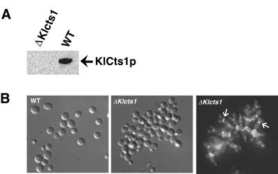
(A) K. lactis Δcts1 cells do not secrete KlCts1p. Proteins in spent culture medium from wild-type GG799 (WT) and Δcts1 K. lactis cells were incubated with chitin beads. Bound proteins were eluted by boiling and were detected by α-ChBD Western blotting. (B) KlCTS1 is required for efficient cell separation. Wild-type and Δcts1 K. lactis cells were grown in YPD medium and fixed in 2.5% glutaraldehyde. Septum-localized chitin was stained with calcofluor white and detected by fluorescence microscopy using a DAPI filter. The middle and right panels show the same cells visualized by phase-contrast and fluorescence microscopy, respectively. The arrows in the right panel indicate the locations of septa in certain cells.

