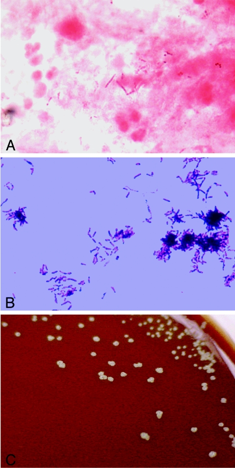
FIG. 3.
Microphotographs and colony morphology of aspirate from the right flank abscess. Panel A shows numerous acid-fast bacilli (Ziehl-Neelsen stain). Panel B shows numerous filamentous, branching gram-positive rods (Gram stain). C shows the typical “molar tooth” appearance of colonies on chocolate agar.

