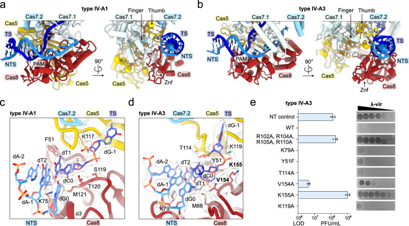
Fig. 2. Cas8 and Cas5 mediate PAM recognition.
a, b View onto the PAM and adjacent dsDNA bound to types IV-A1 (a) and IV-A3 (b) in two 90°-rotated orientations. c and d Close-up view onto the PAM interface in types IV-A1 (c) and IV-A3 (d). Side chains in proximity to the PAM are shown as sticks. The sharpened experimental cryo-EM map is shown as a translucent surface around the side chains and DNA. e λ-vir assay for type IV-A3 probing PAM-interface amino acid substitutions. Plaque Forming Units (PFU) beyond the Limit Of Detection (LOD, countable single plaques) are plotted (blue bars). n = 3 independent spot plate replicates; mean ± s.d. Residues producing interference defects upon substitution are highlighted in bold in panel (d) and Supplementary Fig. 8b. Source data are provided as a Source Data file.

