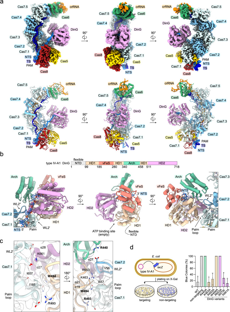
Fig. 6. Cryo-EM structure of type IV-A1 in complex with DinG.
a Top: Sharpened experimental cryo-EM map of the type IV-A1 effector in complex with DinG. Unfiltered and EMReady maps are shown in Supplementary Figs. 2, 4. Below: Structure model of the type IV-A1 effector in complex with DinG. b Top: domain organization scheme of DinG. Below: DinG-centered views. DinG domains are color coded according to the scheme above. c Close-up view onto the Cas7-DinG interface in two 180°-rotated orientations. The sharpened experimental cryo-EM map is shown as a translucent surface. d lacZ-CRISPRi assay probing amino acid substitutions in DinG. The scheme on the left illustrates the assay setup. Coloring in the bar graph according to DinG domain coloring in (b). n = 3 independent replicates; mean ± s.d. Residues producing interference defects upon substitution are highlighted in bold in panel (c). Source data are provided as a Source Data file.

