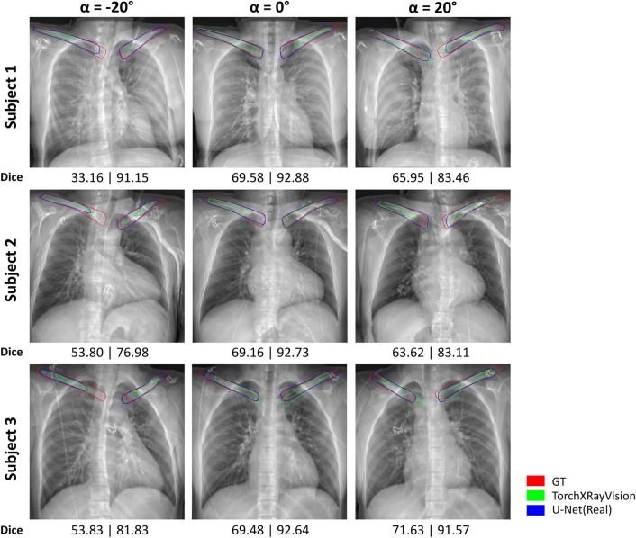
Fig. 3.
Clavicle segmentation results of real-data-trained segmentation models. Three subjects with adversarial-positioned ( and ) and well-positioned () images are shown.The ground truth (GT) segmentation contours from TotalSegmentator are red, TorchXRayVision are green, and U-Net(Real) are blue in color. The Dice score analysis with respect to GT for each patient and angle are shown below each image, left is TorchXRayVision and right is U-Net(Real).

