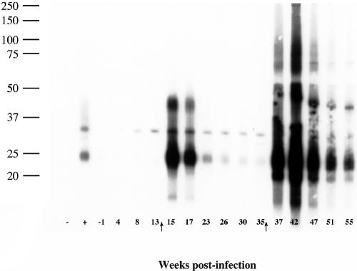
FIG. 2.
Preparative immunoblots of M. bovis strain 95-1315 WCS antigen probed with sera from a reindeer experimentally infected with M. bovis. Molecular mass markers are indicated in the left margin (in kDa) and weeks postinfection in the lower margin. Arrows in the lower margin indicate time points when purified protein derivatives were injected for skin testing (CCT). Symbols in the lower left margin refer to sera from known noninfected (−) and infected (+) white-tailed deer used as controls for the assay. Numbers in the lower margin refer to weeks relative to infection. Immunoblots were performed on samples from each reindeer at all time points indicated. Responses presented in this figure are representative of 10/11 of the infected reindeer. One infected reindeer (animal 125) had only minimal responses detectable by immunoblot analysis.

