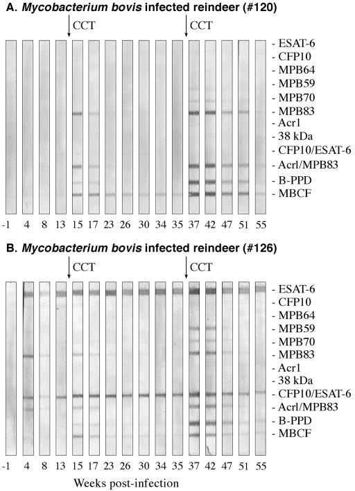
FIG. 3.
Antibody responses to recombinant antigens detected by MAPIA in reindeer experimentally infected with M. bovis. Arrows in the upper margin indicate time points when purified protein derivatives were injected for skin testing (CCT). Antigens printed are shown in the right margin. Strips represent different time points during infection when serum samples were collected (shown in weeks in the lower margin). Representative responses by two different M. bovis-infected animals, animal 120 (A) and animal 126 (B), are provided to demonstrate the variability in antigen recognition patterns.

