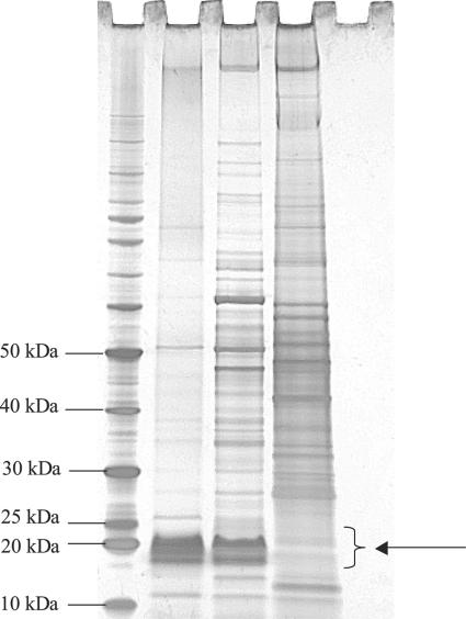
FIG. 1.
SDS-PAGE-periodate silver staining of L. intracellularis LPS. First lane, 10- to 220-kDa benchmark prestained protein marker (Invitrogen); second lane, L. intracellularis LPS extract; third lane, whole-cell L. intracellularis 15540; fourth lane, uninfected McCoy cells; fifth lane, blank. The arrow indicates the migration range of L. intracellularis LPS.

