FIG. 2.
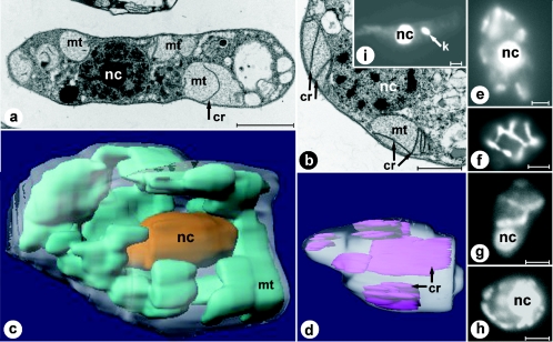
Ultrastructure of D. papillatum mitochondria. Electron microscopy (a and b), light microscopy (e to i), and 3D reconstruction (c and d) based on electron microscopy images. Electron micrographs of longitudinally sectioned (a) and cross-sectioned (b) Diplonema cells. Prominent flat cristae are visible in the organellar lumen. (c) 3D reconstruction of the whole cell, based on 48 consecutive sections. The single mitochondrion is shown in cyan, and the posteriorly located oval nucleus is shown in gold. (d) 3D reconstruction of a large compartment of the mitochondrion, based on 25 consecutive sections. Cristae are shown in purple. (e to g) YOYO1-stained D. papillatum cells focused on the nucleus (e and g) and the cell periphery (f). (h) DAPI-stained D. papillatum cell. (i) Control staining (YOYO1) of the kinetoplastid Trypanosoma brucei. Bars, 2 μm (a), 0.5 μm (b), and 2.5 μm (e to i). nc, nucleus; mt, mitochondrion; k, kinetoplast; cr, cristae.

