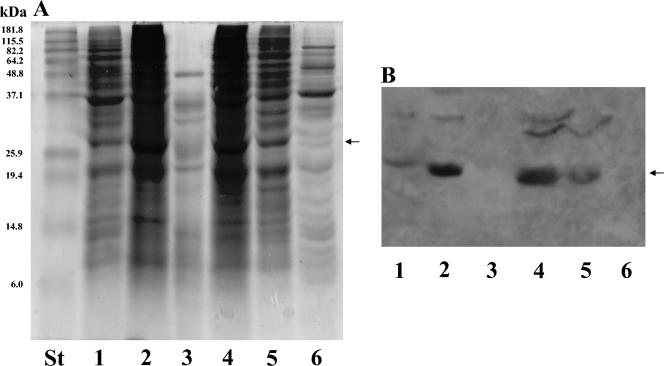
FIG. 2.
SDS-PAGE and Western analysis of subcellular fractions from the C. albicans CAI4 strain. Linear 12% polyacrylamide gels were loaded (50 μg of each sample expressed as total protein content per well) with whole-cell lysate (lane 1), SDS cell wall extract (lane 2), Zymolyase cell wall digest (lane 3), P40 fraction (lane 4), P100 (lane 5), and soluble (cytosol) fraction (lane 6). After electrophoresis, gels were stained with Coomassie blue (panel A) and polypeptides were subsequently transferred to nitrocellulose sheets and immunodetected with PAb anti-Abg1p (panel B). Molecular weight standards were run in parallel with the different samples (lane labeled St in panel A). The arrows point to the band corresponding to Abg1p.

