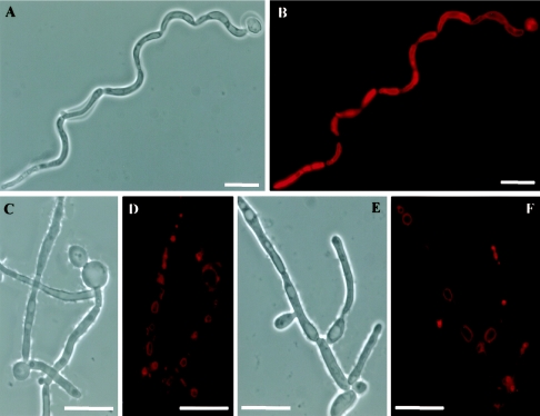
FIG. 8.
Vacuole morphology in hyphal cells. Phase-contrast (A, C, and E) and FM4-64-staining fluorescence observations (B, D, and F) of CAI4-URA3 (A and B) and CV3 (C-F) strains. ABG1 conditional mutant cells had several small vacuoles in each cellular compartment (D, F), while in the wild-type cells a single, large vacuole that occupied almost all the cytoplasmic volume was observed (B). Bar = 10 μm in all panels.

