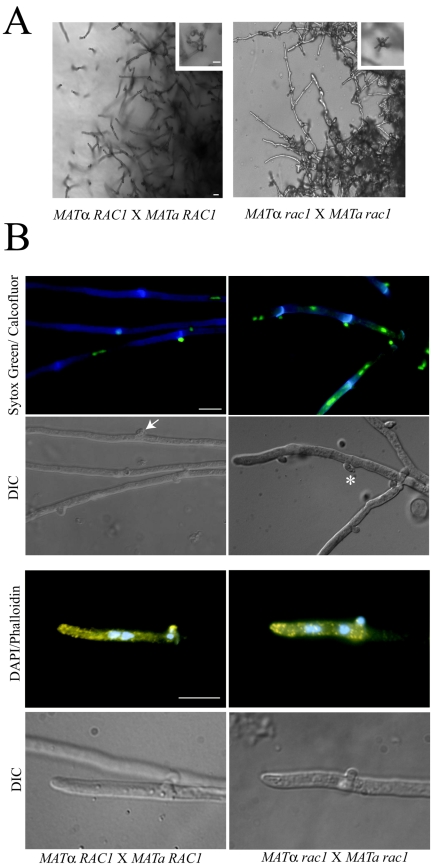
FIG.4.
Altered morphology of the rac1 mutant mating hyphae. (A) Wild-type and bilateral rac1 crosses were incubated on V8 mating medium for 7 days. Areas at the edge of the mating reaction containing filaments were photographed at 200× magnification with DIC optics to visualize filaments, basidia (inset), and basidiospores (inset). Bar, 10 μm. (B) Bilateral wild-type and rac1 crosses were prepared on V8 agar-coated glass slide mounts. After incubation, filaments were fixed and costained with calcofluor and Sytox Green to visualize septa, clamp cell junctions, and DNA (first panel). Filaments and clamp cells were visualized using DIC optics (second panel). Wild-type clamp cells are fused (arrow) while rac1 clamp cells are unfused (*). Wild-type and bilateral rac1 filaments were also costained with rhodamine-conjugated phalloidin and DAPI to visualize actin and DNA (third panel). The fourth panel depicts the corresponding DIC images. Magnification, 1,000×. Bar, 10 μm.

