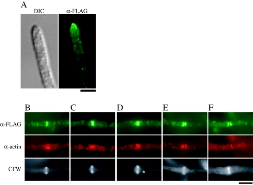
FIG. 7.
Localization of FLAG-ChsC. CΔsC/PG/A/FLAGC-1 was subjected to indirect immunofluorescence using anti-FLAG antibody (α-FLAG) and anti-actin antibody (α-actin). Signals of FLAG-ChsC were observed at the hyphal tips (A) and a subset of the septation sites (B to F). (B to E) Septa with obvious actin and FLAG-ChsC localization. (F) A septum with only FLAG-ChsC localization. Bar, 4 μm.

