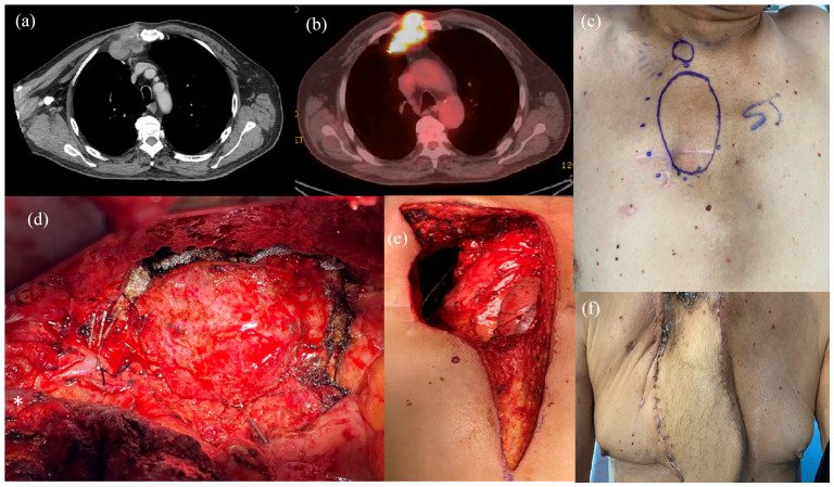
Figure 2.
Re-resection. (a) Regrowth and osteal erosion on 6-month interval CT. (b) Recurrence of avidity on 6-month interval CT PET. (c) Anterior chest wall recurrence with prior resection scar encircled by preoperative plastic surgery markings. (d) Tumor visualized through right posterolateral thoracotomy. Clips are placed on the mammary vessels and branches. Subclavian vein in the lower left-hand corner marked with (*). (e) Postresection anterior chest wall defect with visible mediastinum. The pectoralis muscles were detached from the clavicle to avoid vein injury during the first rib resection, (f) Clinic visit, 1 month postoperatively.

