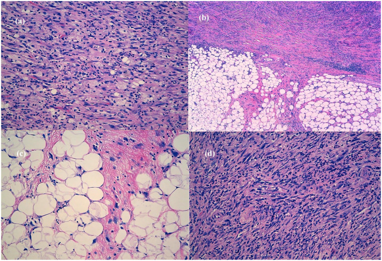
Figure 3.
Final tumor pathology. (a) IMT-like areas: elongated myofibroblasts containing abundant eosinophilic cytoplasm and vesicular nuclei, loose stroma with abundant neutrophils, lymphocytes, and few plasma cells (H&E, 20×), (b) interface between well-differentiated liposarcoma with dedifferentiated foci, well-differentiated liposarcoma with abrupt transition to a nonlipogenic sarcoma (H&E, 5×), (c) well-differentiated liposarcoma component: mature fat with variably sized adipocytes and thick fibrotic bands containing characteristic atypical spindle cells with enlarged, hyperchromatic nuclei. The diagnosis is made by noting the presence of these fibrous bands containing atypical spindle cells (H&E, 20×), (d) cellular high-grade areas of sarcoma component: haphazardly distributed hyperchromatic atypical spindle cells in a fibrous stroma associated mixed inflammatory infiltrate (H&E, 20×).

