Fig. 2.

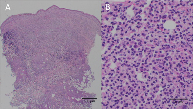
Skin histology from the margin of the wound depicting a deep heavy predominantly neutrophilic infiltrate with admixed histiocytes and some lymphocytes. HE staining: (A) 40x magnification and (B) 400x magnification.

Skin histology from the margin of the wound depicting a deep heavy predominantly neutrophilic infiltrate with admixed histiocytes and some lymphocytes. HE staining: (A) 40x magnification and (B) 400x magnification.