Figure 5.

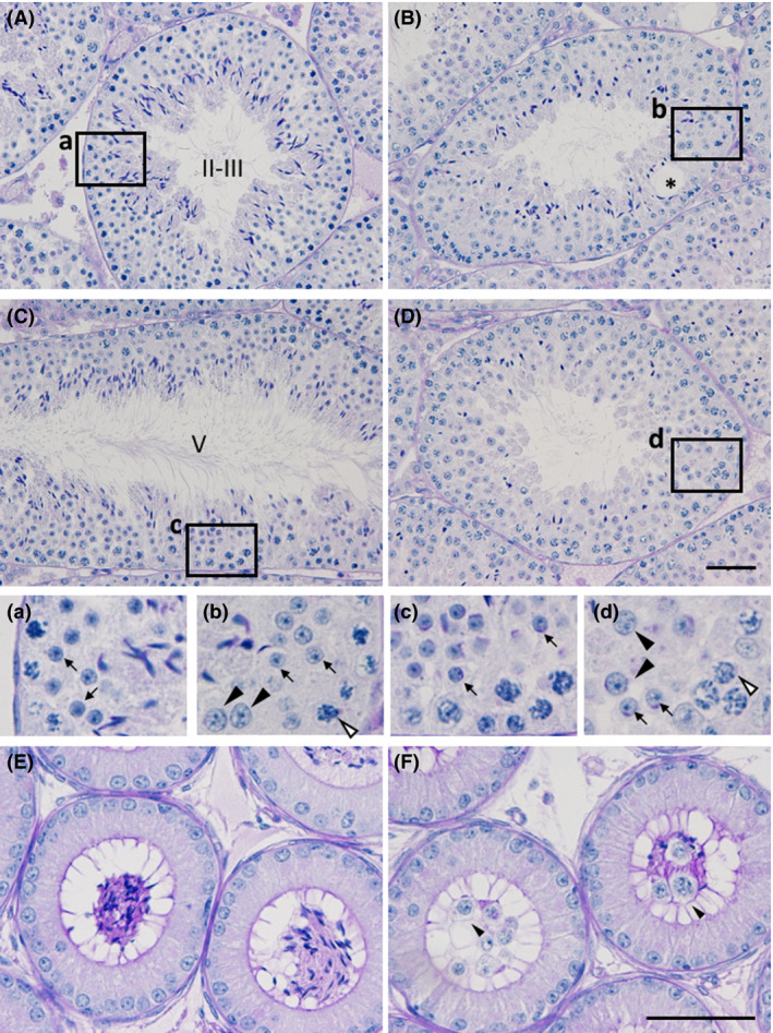
Histological analysis of testes and epididymides of the Zfp318 −/− mouse. PAS‐hematoxylin‐stained sections of testes (A‐D) and caput epididymides (E and F). Depicted areas in A–D are enlarged in a–d, respectively. Roman numerals in A and C show the stage of the cycle of the mouse seminiferous epithelium. Seminiferous tubules in B and D were regarded as the corresponding stage of A and C, respectively. The wild‐type male shows normal testicular morphology (A and C), whereas the mutant testis shows affected spermatogenesis including vacuole formation (B, asterisk) and sparsely located elongated spermatids (D). Note the cells with the large nuclei (b and d, arrowheads) among the normal round spermatids (arrows), comparable to those of pachytene spermatocytes (b and d, white arrowheads). Accumulation of the sperm is seen in the lumen of the wild‐type epididymal duct (E). Sloughed germ cells with few sperm are observed in the mutant (F). Bars: 50 μm.
