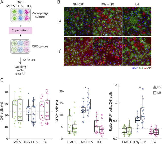
Figure 2. MS Patient Macrophage-Conditioned Media Induce More Astrocytic Differentiation of OPC Than HC Macrophage-Conditioned Media.
(A) Experimental overview for the OPC differentiation assay: HC and MS macrophage-conditioned media were collected after 24 hours of treatment with GM-CSF, IFNγ+LPS, or IL4 and added to cell cultures from the CG4 rat OPC line. (B) Cell fate toward an OL or astrocytic lineage was evaluated using O4 (green, h) and GFAP (red, h) antibodies, respectively. (C) Distribution of percentages of O4+ and GFAP+ cells and the ratio of the 2, grouped by disease group and activation states. The dashed gray line corresponds to the level of O4, GFAP, and the ratio of GFAP/O4 in CG4 cells in absence of macrophage-conditioned media. *p < 0.05, **p < 0.01, ***p < 0.001, and p < 0.0001 in the Mann-Whitney U test between HC and MS for each activation state. For box plots, values are grouped by activation state and disease (HC: triangles; MS: squares; GM-CSF: green; IFNγ+LPS: blue; IL4: pink). HC = healthy controls (n = 14); MS = patients with MS (n = 2).

