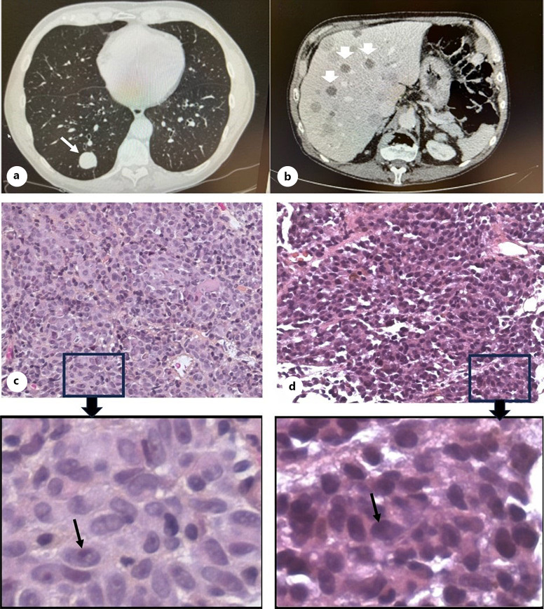
Fig. 3.
a CT scans of lungs in 2016 show one sharply demarcated metastatic lesion in the posterior inferior lobe (white arrow). b CT scans of liver in July 2018 disclose multiple lesions (white arrowheads). Haematoxylin and eosin (HE) staining of metastatic tumour tissues from lung (c) and liver (d) are shown with low (×40) and higher magnification (box around area magnified from ×40). Melanoma spindle B cells with black arrows point to prominent nucleoli of the tumour cells, and pigmentation was only found in some areas.

