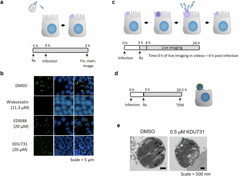
Extended Data Fig. 4. EDI048 does not affect C. parvum sporozoites invasion, parasitophorous vacuole formation or growth but arrests C. parvum at meront stage.
a, Cartoon of life stages investigated in the invasion assay along with schematic of the assay. HCT-8 cells were pre-treated with 2x concentration of compounds for 1 hour (h). Primed C. parvum Iowa strain oocysts were then added and allowed to excyst and invade in presence of 1x compound concentration. At 3 h post-infection parasites that did not invade were washed off and then cells stained for parasitophorous vacuoles using FITC conjuated Vicia villosa lectin (green) and nuclei (blue). The active control wiskostatin (11.3 µM), an inhibitor of N-WASp and its activation by cdc42, prevented sporozoite invasion and formation of trophozoites, whereas 20 µM of EDI048 and KDU731 were inactive (b). c, Graphical representation of stages visualized by time-lapse microscopy and schematic representation of the experimental methodology. HCT-8 cells were infected by oocysts for 3 hours (h) and then washed and treated with 1 µM EDI048 or 0.5 µM KDU731. Time-lapse microscopy images were taken every 20 minutes from 4 h post-infection, that is, 1 h after compound addition (time 0 h in the videos) up to 24 h post-infection and are shown in Supplementary Videos 1 (DMSO), 2 (EDI048), and 3 (KDU731). d, Experimental overview to determine the effect of KDU731 on meronts by transmission electron microscopy (TEM). HCT-8 cells were infected with C. parvum oocysts induced for excystation and 0.5 µM of KDU731 was added at 3 h post-infection and parasite morphology analyzed by TEM at 10.5 h post-infection (e). Scale bars: 5 µm (b and Supplementary Videos 1 to 3) and 500 nm (e). Images and videos are representative of at least 2 independent experiments.

