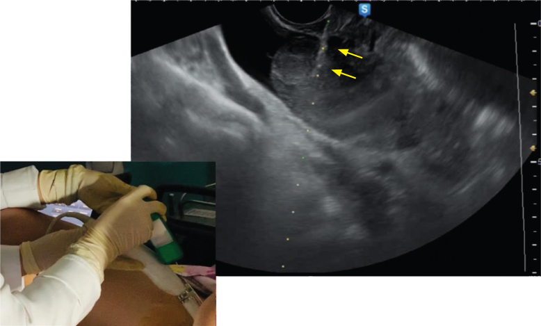
Figure 4.
Tru-cut biopsy of the right ovarian mass done under transvaginal ultrasound guidance. The tru-cut needle is inserted through the needle guide attached to the transvaginal probe. After securing placement of the needle within the tumor, the biopsy gun is fired. The biopsy needle (yellow arrows) is shown penetrating into the lesion. The dotted line represents the track of the needle.

