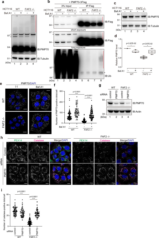
Fig. 4. Extraction of excess PMP70 by the p97/VCP-FAF2 complex prevents pexophagy.
a WT or FAF2-/- HCT116 cells were treated with or without Baf.A1 for 24 h and immunoblotted with the indicated antibodies. The red vertical line denotes a smear in the PMP70 signal. The asterisks indicate cross-reactive bands. n = 3 assays. b PMP70 ubiquitylation in FAF2-/- cells. WT or FAF2-/- cells stably expressing PMP70-3Flag were treated with or without Baf.A1 for 24 h and then immunoprecipitated with anti-Flag antibodies. The samples were immunoblotted with anti-Flag and anti-ubiquitin antibodies. The red vertical lines denote ubiquitylation. The asterisks indicate cross-reactive bands. n = 2 assays. c PMP70 level in FAF2-/- cells increases in response to Baf.A1 treatment. The cells were treated with or without Baf.A1 for 24 h and then immunoblotted with the indicated antibodies. d Quantitative analysis of PMP70 levels in cells from c. PMP70 levels were normalized to WT cells, which were set to 1. Dots indicate individual data points from three independent experiments. Statistical significance was calculated using a one-tailed Welch’s t-test; *p < 0.05. The center lines correspond to the medians and the box limits indicate the 25th and 75th percentiles. The box-plot whiskers extend 1.5 times the interquartile range from the 25th and 75th percentiles. e Peroxisomal abundance in FAF2-/- cells is recovered following Baf.A1 treatment. At 24 h post-Baf.A1 treatment, the cells were immunostained with anti-PMP70 antibodies. Cell nuclei were stained with DAPI. Scale bars, 10 µm. f Quantitative analysis of the per cell number of PMP70-positive peroxisomes for cells in e. The number of PMP70-positive peroxisomes per cell was plotted. Dots represent individual data points from three independent experiments. n = 103 cells (35, 35, 33 cells/experiments; WT), n = 68 cells (22, 22, 24 cells/experiments; WT + Baf.A1), n = 91 cells (32, 30, 29 cells/experiments; FAF2-/-), and n = 110 cells (37, 37, 36 cells/experiments; FAF2-/- + Baf.A1). Bars, median. Statistical significance was calculated using one-way ANOVA; ****p < 0.0001. g WT and FAF2-/- cells were treated with control or PMP70 siRNAs and then immunoblotted with the indicated antibodies. h PMP70 knockdown reverted the peroxisomal loss observed in FAF2-/- cells. WT and FAF2-/- cells were treated with control or PMP70 siRNAs and then immunostained with anti-PEX14 and anti-catalase antibodies. Cell nuclei were stained with DAPI. Scale bars, 10 µm. i Quantitative analysis of the per cell number of catalase-positive peroxisomes per cells in h. The dots indicate individual data points from three independent experiments. n = 86 cells (35, 26, 25 cells/experiments; WT + sicontrol), n = 73 cells (23, 26, 24 cells/experiments; WT + siPMP70, n = 100 cells (33, 34, 33 cells/experiments; FAF2-/- + sicontrol), and n = 73 cells (25, 25, 23 cells/experiments; FAF2-/- cells + siPMP70). Bars, median. Statistical significance was calculated using one-way ANOVA; ****p < 0.0001. Source data are provided as a Source Data file.

