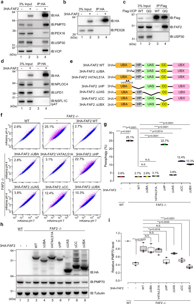
Fig. 5. FAF2 acts as a negative regulator of basal pexophagy.
a, b HCT116 cells stably expressing 3HA-FAF2 were treated with the chemical crosslinker DSP and then co-immunoprecipitated with anti-HA agarose beads. The samples were immunoblotted with the indicated antibodies. 3HA-FAF2 interacts with PMP70, PEX16, USP30, and p97/VCP, but not PEX26. n = 3 assays. c p97/VCP interacts with FAF2 but not USP30. HeLa cells transiently expressing Flag-tagged p97/VCP WT or the E305Q/E578Q mutant (QQ) were treated with DSP and then co-immunoprecipitated with anti-Flag beads. The samples were immunoblotted with the indicated antibodies. n = 2 assays. d 3HA-FAF2 forms a complex with NPLOC4 and UFD1 but not NSFL1C/p47. HCT116 cells stably expressing 3HA-FAF2 were treated with DSP and then immunoprecipitated with anti-HA agarose beads. The samples were immunoblotted with the indicated antibodies. n = 2 assays. e Schematic diagram of the FAF2 constructs used in this study. UBA, ubiquitin-associated domain; HP, hairpin; CC, coiled-coil domain; UBX, ubiquitin-regulatory X domain. To disrupt interactions with ubiquitin, V47A and L51A substitution were introduced into the UBA domain. deletion; Δ. f FACS-based analysis of the pexophagy flux. Representative FACS data (mKeima-SKL 561/488 ratio) for WT, FAF2-/-, or FAF2-/- cells stably expressing the indicated 3HA-FAF2 mutants with the percentage of pexophagy-positive cells indicated. FAF2 ΔUAS, FAF2 ΔUBA, and FAF2 V47A/L51A restored pexophagy in FAF2-/- cells. The effect of pexophagy suppression by FAF2 ΔHP, ΔCC, and ΔUBX was limited, indicating that the FAF2 UBA and UAS domains are dispensable for pexophagy suppression. deletion; Δ. g Quantitative analysis of the pexophagy flux in f. Dots represent individual data points from three independent experiments. Statistical significance was calculated using one-way ANOVA; **p < 0.01; **** p < 0.0001; N.S.—not significant. The center lines correspond to the medians, and the box limits indicate the 25th and 75th percentiles. The box-plot whiskers extend 1.5 times the interquartile range from the 25th and 75th percentiles. deletion; Δ. h Total cell lysates prepared from WT, FAF2-/-, and FAF2-/- HCT116 cells expressing 3HA-FAF2 WT or the indicated mutants were immunoblotted with the indicated antibodies. deletion; Δ. i Quantitative analysis of PMP70 levels in cells from h. PMP70 levels were normalized to WT HCT116 cells, which were set to 1. Dots indicate individual data points from three independent experiments. Statistical significance was calculated using one-way ANOVA; **p < 0.001; ***p < 0.001; ****p < 0.0001; N.S. not significant. The center lines correspond to the medians, and the box limits indicate the 25th and 75th percentiles. The box-plot whiskers extend 1.5 times the interquartile range from the 25th and 75th percentiles. deletion; Δ. Source data are provided as a Source Data file.

