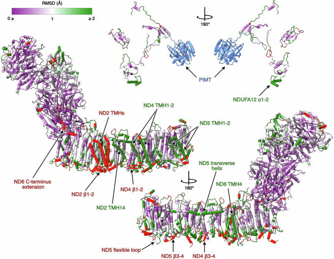
Fig. 2. Cα-RMSD comparison of the structures of P. denitrificans and B. taurus (PDB-7QSL20) complex I.
The structure of Pd-CI is coloured by Cα RMSD value following per-subunit alignment in UCSF ChimeraX. A purple-white-green palette from 0 to 2 Å was used to colour the structure. Unpaired regions specific to P. denitrificans complex I are in red and the P. denitrificans-specific PIMT subunit is in blue. The core and supernumerary subunits are shown separately with key elements of interest indicated. See Fig. 1 for the locations of individual subunits.

