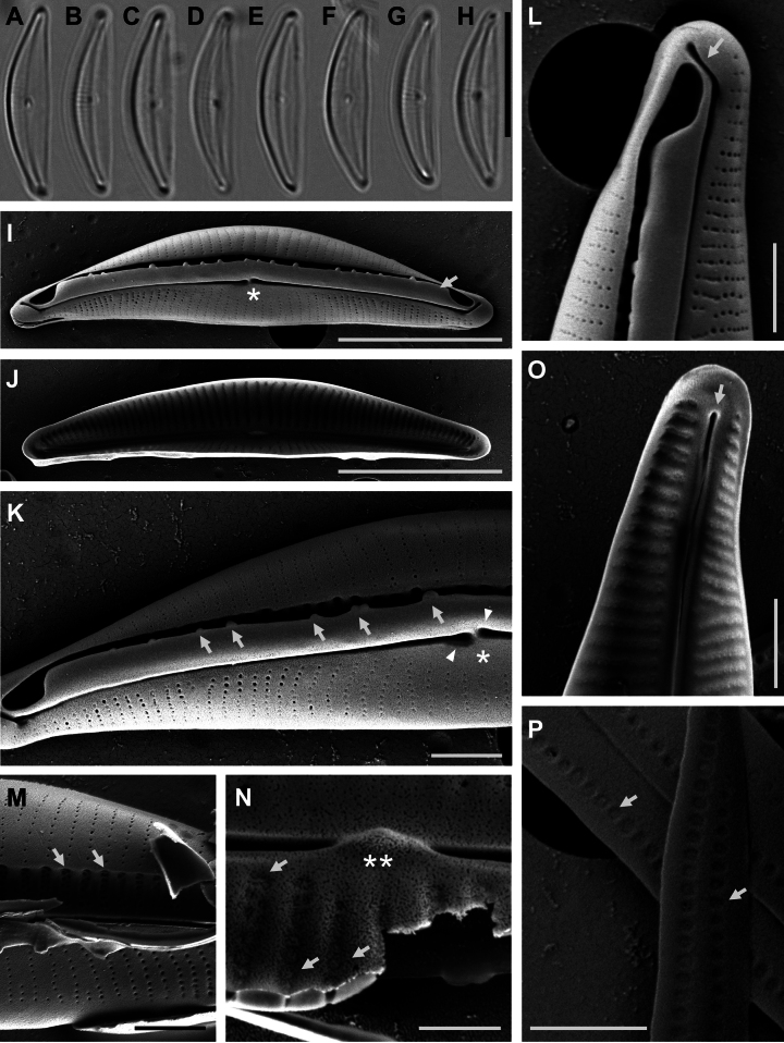
Figure 2.
A–H Light microscopy photomicrographs of H.hampyeongensisI–P scanning electron microscopy photomicrographs of H.hampyeongensisI external valve view, with central area (asterisk) and dorsal raphe ledge (arrow) J internal view of a valve K detail of a valve externally showing siliceous outgrowths (arrows) on the margin of the raphe ledge, central area (asterisk), and proximal raphe endings (arrowheads) L detail of external valve apex showing the dorsally bent distal raphe ending (arrow) M biseriate striae (arrows) in several rows under the raphe ledge N detail of areolae on the dorsal side internally occluded by hymenes (arrows) and tongue-like proximal helictoglossae (double asterisk) O detail of internal valve apex showing poorly developed distal helictoglossae (arrow) P girdle bands with two rows of poroids (arrows). Scale bars: 10 μm (A–H); 5 μm (I, J); 1 μm (K–M, O, P); 0.5 μm (N).

