
Figure 1.
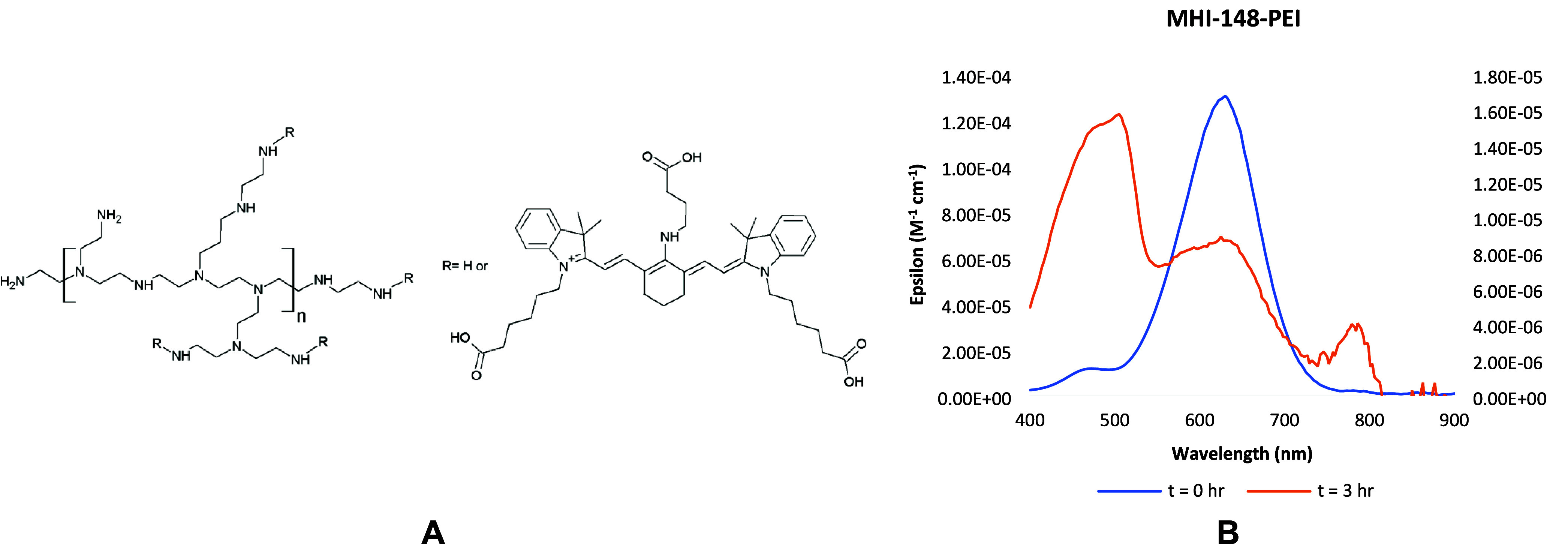
A. Structure of MHI-148-PEI;17 B. Absorption spectra of MHI-148-PEI with a peak at 465 nm coming from the partially degraded dye at t = 0 h (in blue corresponding to the y-axis on the left) and at the end of 3 h (in orange corresponding to the y-axis on the right) of continuous illumination.