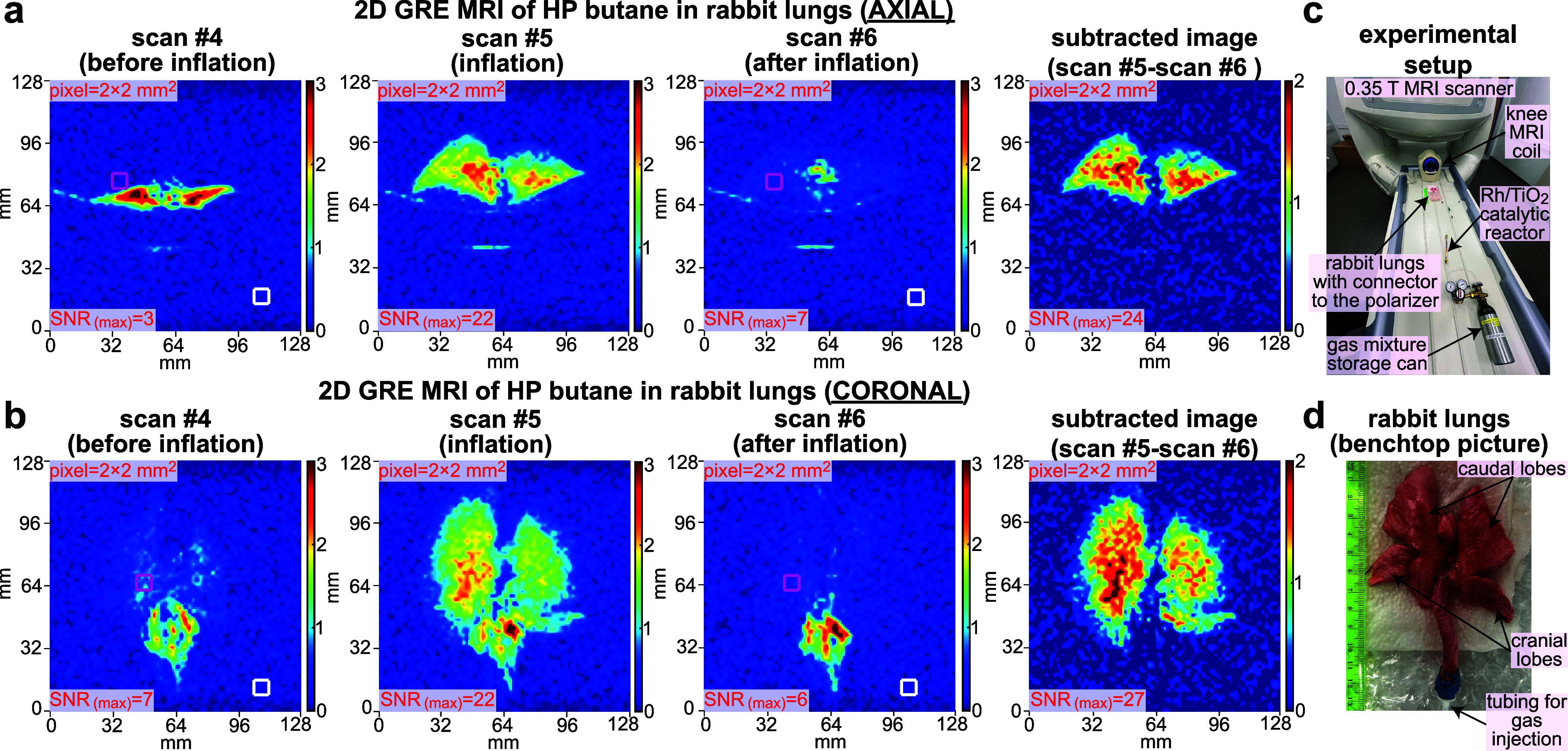
Figure 5.

Subsecond 2D ventilation MRI using HP butane gas contrast agent injected in the excised rabbit lungs. (a) Time series of axial scans recorded before lungs’ inflation with the HP butane (scan #4), at inflation (scan #5), after the inflation (scan #6), and the difference (or “subtracted”) image obtained by subtracting the “scan #6” from “scan #5” image (to remove the background signals from surrounding tissues). The imaging parameters employed were the same as those used in Figure 4 except for the FOV that was reduced to 128 × 128 mm2. (b) Corresponding time series recorded on a different HP butane gas injection in the same lungs in the coronal projection. (c) Annotated photo of experimental setup showing the gas mixture storage tank, Rh/TiO2 reactor, 0.35 T knee MRI coil, and gas connection from the reactor outlet to the excised rabbit lungs. (d) Annotated photograph of the excised rabbit lungs used in the study.
