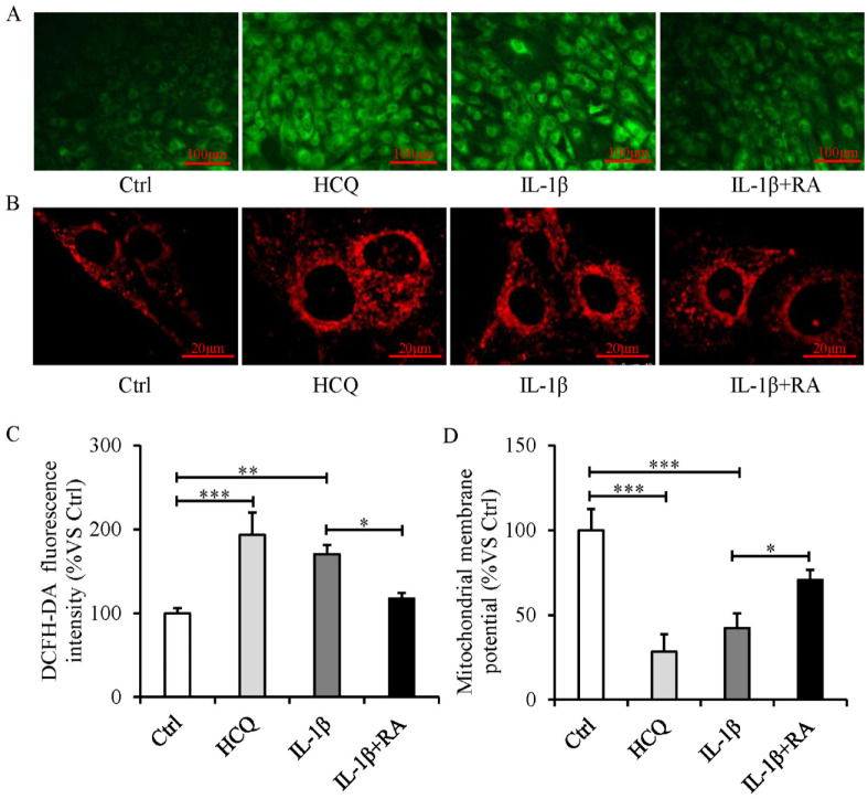
Figure 5.
Mitochondrial dysfunction and intracellular ROS elevation occur during autophagy defect, and autophagy activation attenuates mitochondrial dysfunction and decreases ROS production in IL-1β-treated SFZCs. SFZCs were treated with HCQ (10 μM), RA (100 nM), or IL-1β (20 ng/ml) for 5 days. (A) ROS assessment using DCFH-DA fluorescence. (B) Mitochondrial staining using MitoTracker. (C) DCFH-DA fluorescence quantified using flow cytometry. (D) Mitochondrial membrane potentials were measured using JC-1 staining, expressed as the ratio of red to green fluorescence. ROS = reactive oxygen species; IL-1β = interleukin-1β; SFZCs = superficial zone cells in articular cartilage; HCQ = hydroxychloroquine; RA = rapamycin; DCFH-DA = 2’-7’dichlorofluorescin diacetate. *P < 0.05, **P < 0.01, and ***P < 0.001.

