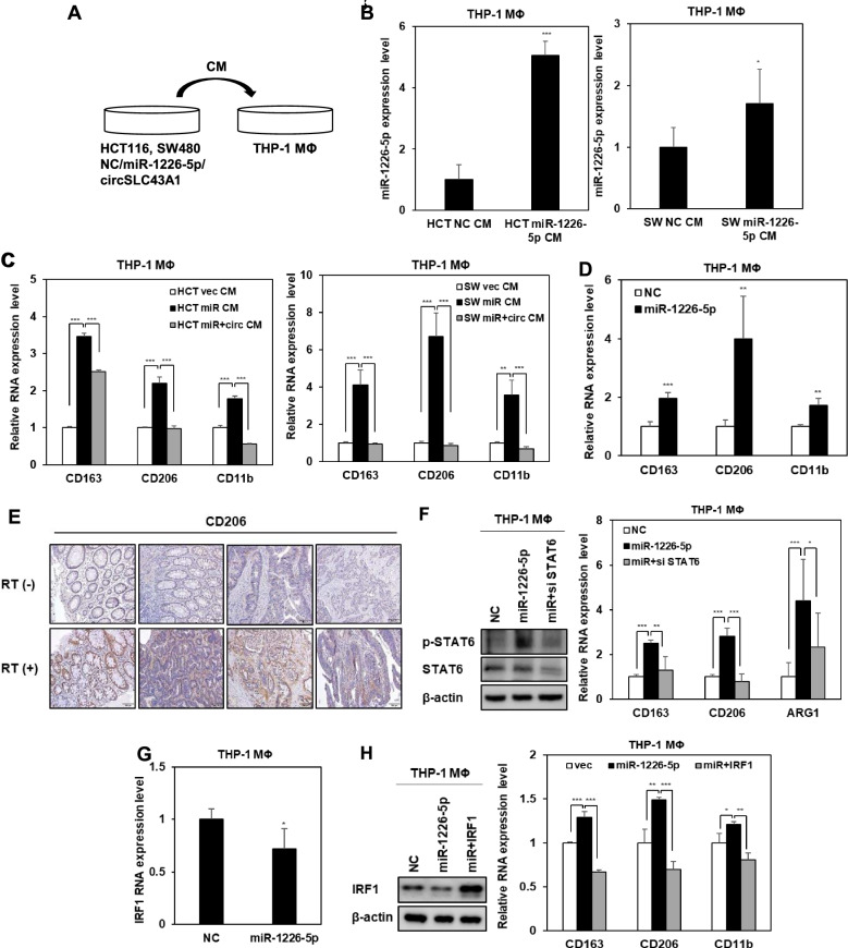
Fig. 4.
miR-1226-5p induces M2 polarization via IRF1 inhibition in THP-1-derived macrophages. A–D Human monocytes, THP-1, were differentiated into macrophages by treating them with PMA (100 ng/ml) for 24 h. A Conditioned media harvested from HCT116 and SW480 overexpressing either miR-1226-5p and circSLC43A1 were treated with THP-1-derived macrophages. B The level of miR-1226-5p in the indicated THP-1-derived macrophages was confirmed by qRT-PCR. C The mRNA levels of CD163, CD206, and CD11b were measured in the indicated THP-1-derived macrophages. D After overexpression of the miR-1226-5p mimic in THP-1-derived macrophages, the expression of CD163, CD206, and CD11b was confirmed by qRT-PCR. E The expression of CD206 in tissues of CRC patients with and without radiotherapy was compared by IHC. Scale bar 100 um. F After co-transfection of siRNA against STAT6 and miR-1226-5p in THP-1-derived macrophages, protein expression of p-STAT6, and STAT6 was compared through Western blot analysis (left). The mRNA levels of CD163, CD206, and ARG1 were measured in the indicated cells (right). G The mRNA expression of IRF1 upon overexpression of miR-1226-5p in THP-1-derived macrophages was analyzed by qRT-PCR. H After co-transfecting THP-1-derived macrophages with miR-1226-5p and IRF1 overexpression vector, the protein expression of IRF1 was compared through Western blot analysis (left). The mRNA levels of CD163, CD206, and CD11b were measured in the indicated cells (right). qRT-PCR was normalized to GAPDH and U6. β-Actin was used for normalization in Western blot analysis. The experiment was repeated three times and representative Western blot images are shown. The data are presented as the mean ± S.D. *P < 0.05; **P < 0.01; ***P < 0.001. Student’s t test, and One-way ANOVA followed by bonferroni comparison test

