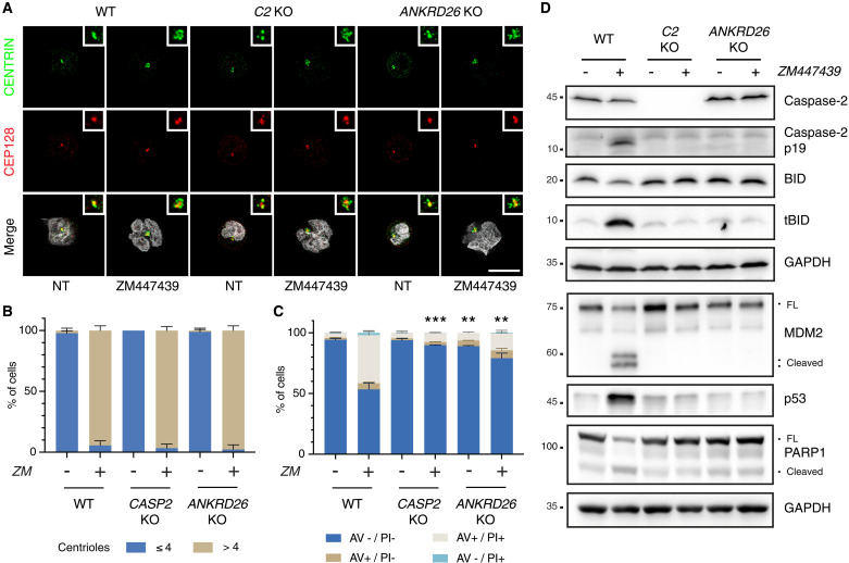
Fig. 7. Recruitment of PIDD1 to the centrosome via ANKRD26 is necessary for cytokinesis failure–dependent cell death.
(A) Representative immunofluorescence of Nalm6 WT, caspase-2, and ANKRD26 KO derivative clones untreated or treated with 2 μM ZM for 48 hours. Cells were costained with the indicated antibodies: CENTRIN (in green) and CEP128 (in red). Hoechst was used to visualize the DNA (gray in the merge). Scale bar, 5 μm. (B) Quantification of the number of centrioles of cells shown in (A). Data are presented as means ± SD (in percentage) of three independent biological replicates. For each replicate and condition, 30 cells were counted. (C) Percentage of Nalm6 WT, caspase-2 KO, and ANKRD26 KO derivative clones undergoing apoptosis after 48 hours of treatment with 2 μM ZM as detected by annexin V/PI staining and flow cytometric analysis. Data are presented as means ± SD of events in each staining condition (in percentage) of N = 3 independent biological replicates. Statistics were calculated by unpaired t test comparing the percentage of live cells of each KO clone to the corresponding treatment condition in the WT sample. **P < 0.01; ***P < 0.001. (D) Western blot showing Nalm6 WT cells or clones edited for caspase-2 or ANKRD26 after 48 hours of treatment with 2 μM ZM.

